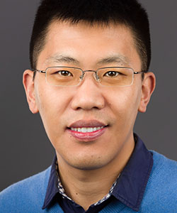
Assistant Professor
Department of Biological Sciences, College of Science
Dr. Chang arrived in January 2018 and is the newest recruit to the Purdue molecular biophysics community. As a post-doctoral fellow, he used cryo-EM to determine high-resolution structures of protein complexes such as the SNARE recycling complex, the cyanobacterial light-harvesting complex, and the anaphase-promoting complex. Currently his lab is interested in the molecular mechanisms of cell cycle regulation. Cryo-EM single particle reconstruction will be a major focus for trainees.
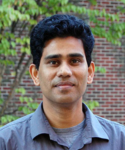
Professor
Department of Chemistry, College of Science
The Das group relies on X-ray crystallography for structural characterization of deubiquitinases (DUBs) and their complexes with interacting partners. For validation they use biophysical methods to investigate protein-protein interactions in solution. Isothermal calorimetry (ITC) is used to determine affinity parameters of DUB variants for their binding partners. Graduate students are trained on the use of ITC instruments administered by the director of the Bindley Biophysical Analysis Laboratory, as well as data analysis using software such as SEDPHAT. Similar training applies to analytical ultracentrifugation experiments for quantitative determination of protein-protein interaction parameters.
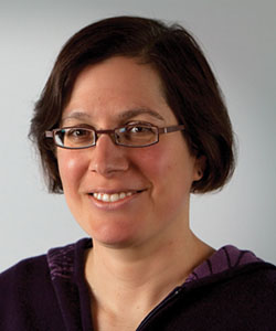
Professor
Department of Biochemistry, College of Agriculture
Non-coding RNA sequences are very important for regulation of gene expression and other cellular processes, and many fold into stable three-dimensional structures in order to function. A wide variety of riboswitches and catalytic RNAs continue to be discovered in a variety of contexts, including eukaryotic transcriptomes. A detailed molecular understanding of these systems is required to understand their catalytic and regulatory functions. Trainees use crystallographic and mechanistic analyses of functional RNAs with the goal of obtaining high-resolution snapshots of how these molecules work within the cell.
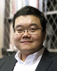
Associate Professor
Department of Biomedical Engineering, College of Engineering
Dr. Huang brings expertise in high-resolution molecular imaging of cell, viruses and tissues. His lab develops super-resolution imaging techniques such as single molecule switching nanoscopy (SMSN, also known as PALM/STORM). To push the limits of nanoscopy towards imaging cellular dynamics and deep tissues, the lab develops novel nanoscopy methods using, for example, adaptive optics, single molecule interferometry nanoscopy (4PiSMSN), and massively parallelized computing. Through extensive collaboration with Purdue and the IU School of Medicine, trainees apply these novel capabilities to resolve structure, dynamics and function of cells/tissues under drugged, toxic, wounded, and physiological conditions.
Professor
Department of Biological Sciences, College of Science
Dr. Jiang uses cryo-EM to study the structures of viruses, large macromolecular complexes, and membrane proteins. Trainees also work on development of various cryo-EM methodologies including image processing algorithms, high performance computing, data collection automation, and sample preparation. These technical advances will help cryo-EM single particle and tomographic 3-D reconstructions to be routinely solved at near atomic resolutions, and ultimately evolve these techniques into easily accessible and high-throughput tools.
Professor
Departments of Biological Sciences and Computer Science, College of Science
Dr. Kihara's research projects include development and application of computational methods for protein-protein docking, protein structure prediction, protein structure modeling for electron microscopy density maps, virtual drug screening, and protein function prediction. Students are trained in computational skills including coding and the use of state-of-the-art cluster computers and GPUs, and in the biophysical principles of protein folding and ligand-protein interactions. Students learn teamwork through collaboration with experimental labs and also gain supervising and teaching skill by working with undergraduate students and providing guest lectures in the bioinformatics courses that Kihara instructs.
Associate Professor
Department of Biomedical Engineering, College of Engineering
Research projects in the Kinzer-Ursem lab are focused on quantitative characterization of spatial and temporal parameters that affect protein network behavior. Trainees utilize a multidisciplinary approach combining experimental and computational tools to describe structural, functional and regulatory relationships among proteins that are important regulators of excitatory cell behavior, particularly synaptic plasticity in neurons. The Kinzer-Ursem Lab uses high resolution imaging techniques (cryo-EM and super-resolution microscopy) and continuous and stochastic mathematical modeling to describe protein function. We also utilize in vivo protein tagging techniques to isolate and identify newly synthesized proteins. Ultimately, these techniques are combined with systems biology models of protein network signaling to quantitatively describe the molecular mechanisms that underlie cellular behavior in normal and disease states.

Distinguished Professor
Director of Purdue Institute of Inflammation, Immunology and Infectious Disease (PI4D)
Department of Biological Sciences, College of Science
The Kuhn laboratory studies the molecular mechanisms involved in viral replication and pathogenesis, focusing on several groups of RNA-containing viruses that include significant human pathogens. They study the assembly of the virus particle, perform molecular analyses of structural proteins and their role in pathogenesis, and investigate the replication of positive-strand RNA genomes and interactions with the host. In each area they use a combination of biochemical, molecular genetic and biophysical techniques to gain insight into the molecular requirements for the replication, infectivity, and pathogenesis of the virus. Trainees are currently involved in collaborative research and joint training of graduate students in conjunction with several other mentors of this training grant.
Assistant Professor
Department of Biochemistry, College of Agriculture
Understanding the enzymology and genetics of specialized metabolism is a first step towards engineering organisms to produce valuable antibiotics and commodity chemicals. However, a missing link in the engineering process is an understanding of the basic biophysical properties of enzymes. For example, how do the enzymes use binding energy or the excess energy from the reactions they catalyze to alter the overall catalytic rates, and how does mutating those enzymes alter substrate specificity or affect those rate enhancement strategies? Trainees use synthetic organic chemistry to generate transition-state or intermediate analogs for the study of reaction pathways in enzymes by structural biology, especially X-ray crystallography.

Distinguished Professor
Department of Chemistry, College of Science
Dr. Low's trainees have developed small-molecule conjugates that can be targeted specifically to pathologic cells while avoiding collateral toxicity to healthy cells. In the case of cancer, the lab has exploited upregulation of high-affinity folate receptors on carcinomas found in ovary, breast, endometrium, lung, and kidney, and designed imaging and therapeutic agents that target these cancers. More recently, Low has developed a targeting ligand that selectively delivers attached drugs to prostate cancer cells, enhancing both the diagnosis and treatment of prostate cancer. Ligand-directed imaging and drug delivery for multiple autoimmune, inflammatory, and infectious diseases are also being developed. Low has graduated 62 PhD students including 5 under-represented minorities (URMs), with approximately half employed in academia and research hospitals, and the remainder pursuing careers in the pharmaceutical industries. Low and trainees have helped to found several companies in order to commercialize their advances, including Endocyte (publicly traded on NASDAQ), HuLow, and On-Target.
Assistant Professor
Departments of Chemistry & Physics and Astronomy, College of Science
Many important signaling reactions are initiated at the interface of two cells. The Low-Nam lab employs high spatiotemporal resolution imaging coupled with reconstitution strategies to directly visualize cellular signal transduction. These techniques enable a mechanistic characterization of the roles of molecular binding events, protein assembly, membrane topography, and environmental mechanics to signaling within the plasma membrane reaction landscape. The Low-Nam group focuses on quantification of signaling dynamics and heterogeneity in single immune or tumor cells, whose cell-cell interactions have important implications for cancer immunotherapy. Trainees will be exposed to a highly interdisciplinary environment using approaches in fluorescence microscopy, image processing, cellular biophysics, and membrane physical chemistry.

Associate Professor
Departments of Biological Sciences and Chemistry, College of Science
Phospholipase C (PLC) β and ε enzymes are critical regulators of intracellular Ca2+ and protein kinase C activity in the cardiovascular system. However, the molecular mechanisms governing their regulation under normal and pathologic states is poorly understood. The Lyon Lab uses X-ray crystallography, small angle X-ray scattering, electron microscopy, and functional assays to elucidate the mechanisms regulating these enzymes. Trainees in the lab have the opportunity to master cutting edge techniques in these areas, as well as learn how to make their experiments robust, rigorous, and applicable to human health.
Associate Professor
Department of Biological Sciences, College of Science
Trainees employ a multidisciplinary approach to understanding the functional repertoire of Fic (filamentation induced by cyclic-AMP) proteins in regulating prokaryotic and eukaryotic signal transduction pathways, including X-ray crystallography. Fic proteins carry out diverse post-translational modifications, predominantly adenylylation/AMPylation, which involves the covalent addition of adenosine monophosphate to a target protein. Most recently, trainees in her laboratory identified a hitherto unknown role for the sole human Fic protein, HYPE/FicD, in regulating the mammalian unfolded protein response by adenylylating the ER chaperone BiP. They have now extended these findings to diseases attributed to protein misfolding.

Professor, Department Head
Department of Biochemistry, College of Agriculture
Dr. Mesecar investigates the structure and function of biomedically important enzymes using X-ray crystallography, enzyme chemistry & kinetics, proteomics, assay development and optimization, high-throughput screening, and molecular modeling. Mesecar has pioneered the structure-based design of highly selective, non-covalent inhibitors of deubiquitinating enzymes such as papain-like protease (PLPro) in the SARS virus, and ubiquitin-specific protease-7 (USP7), which regulates the P53/MDM2 axis in cancer. Other research projects address the structure and function of the Keap1-Cul3-Rbx1 Ubiquitin E3-ligase system, the mechanisms of bacterial adenylyltransferases in the CoA, NAD, FAD and menaquinone biosynthesis pathways, and the structure-based design of targeting ligands against Muc1, an oncoprotein expressed by 90% of known cancers.
Assistant Professor
Department of Biological Sciences, Joint Appointment in Department of Chemistry, College of Science
Dr. Metskas arrived at Purdue in August 2021. Projects in the Metskas lab currently focus on two main areas: the polymerization and lattice formation of enzymes in bacterial microcompartments responsible for carbon fixation, and the arrangement of flavivirus glycoproteins during membrane fusion and cell entry. In both areas, the arrangement of proteins within the assemblies affect their collective function, and the Metskas lab is dedicated to determining the ultrastructure-function relationship in these systems. We use a combination of cryo-electron tomography (structure), fluorescence spectroscopy (function), and correlated light and electron microscopy.
Associate Professor
Department of Biological Sciences, College of Science
The Noinaj lab is interested in understanding how pathogenic bacteria use virulence factors found on their membranes to mediate infection. These virulence factors are present in the outer membrane of Gram-negative bacteria or within plastids such as in malaria and Toxoplasma gondii. In particular, the lab studies two essential membrane-embedded complexes: the BAM complex from E. coli and Neisseria and the TOC complex from plastids. Trainees use structural analysis by X-ray crystallography/low-angle scattering/EPR/EM and functional studies of these assemblies with the long-term goal of drug discovery and development against multidrug resistance pathogens.
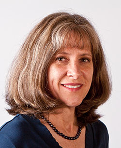
Distinguished Professor
Department of Medicinal Chemistry and Molecular Pharmacology, College of Pharmacy
The Post lab studies the structural, dynamical, and energetic properties of biological molecules and molecular recognition associated with ligand binding, protein-protein interactions, enzymatic catalysis, and conformational equilibrium. Recent goals are to describe and understand the mechanism of conformational activation of tyrosine kinases, the structural response to tyrosine phosphorylation in signal transduction, and large-scale concerted dynamics of viral capsids as it relates to antiviral mechanisms of action and capsid molecular recognition. Trainees learn a variety of biophysical techniques with emphasis on solution NMR, and utilize and develop computer molecular dynamics simulation methods, a natural complement to NMR. Post is one of the developers of the molecular dynamics program CHARMM.
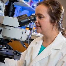
Professor
Department of Physics and Astronomy, College of Science
Dr. Pushkar focuses on the function of transition metals at different length scales from molecules of purified metalloproteins to brain tissues and single cells. Her extensive background in X-ray spectroscopy, imaging, and EPR provides her trainees with research tools of unprecedented sensitivity to generate a wealth of information regarding the oxidation states and coordination environments of metals in diluted systems such as metalloproteins and brain cells. Training the next generation of scientists capable of utilizing complex physical techniques, time resolved and in situ spectroscopies for research in biophysics is also an important part of Dr. Pushkar's research activity, with an interdisciplinary approach key to obtaining new discoveries and to future scientific progress.
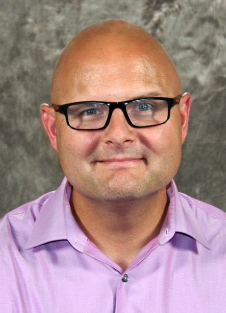
Professor
Department of Medicinal Chemistry and Molecular Pharmacology, College of Pharmacy
Dr. Stahelin’s research focuses on the molecular basis of lipid-protein interactions. The major systems under investigation are virus structure and assembly from the host cell plasma membrane and sphingolipid signaling in inflammation and cancer. Biophysical studies in vitroand in cell culture of protein interactions at the membrane interface utilize methods such as single molecule imaging, super resolution microscopy, and structural biology (cryo-EM). Complementary biophysical techniques such as surface plasmon resonance, isothermal titration calorimetry, hydrogen-deuterium exchange mass spectrometry, and fluorescence are used to gain a mechanistic understanding of the molecular basis of lipid binding and protein conformational dynamics in the presence of membranes.
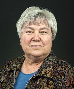
Professor
Department of Biological Sciences, College of Science
Her lab applies X-ray crystallography and molecular biology to systems where proteins work together in membranes to perform biological functions in cancer, heart disease, and transport molecule-related diseases. Structural and functional studies rely on combinatorial approaches, such as crystallographic and EPR investigation of ABC transporters from bacteria and mammals. Mechanistic studies of the enzymes involved in isoprenoid biosynthesis in mammals and pathogenic bacteria have expanded into time-resolved X-ray crystallography and antibacterial drug design. Phosphatase and kinase partners critical in the cell cycle and in the development of metastatic potential in cancer are a third topic of investigation. Structural studies, combined with analytical ultracentrifugation, mass spectrometry, surface plasmon resonance, and fluorescence spectroscopy give a multifaceted biophysical view of these complex systems.
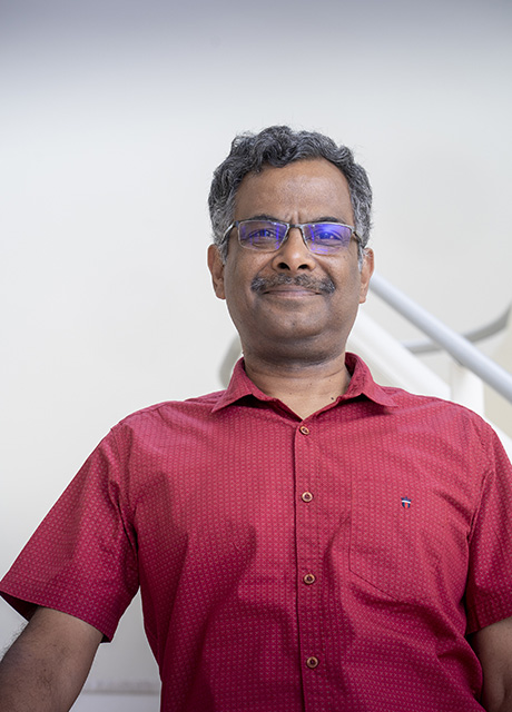
Ramaswamy Subramanian (AKA Rams)
Professor and Director of Bindley Bioscience Center
Department of Biological Sciences, College of Sciences & Department of Biomedical Engineering, College of Engineering
Rams arrived at Purdue summer of 2019 after working at Uppsala, Iowa City, and Bangalore. The Lab uses X-ray crystallography, cryo-EM, and other biophysical tools to study structure function relationships of biomolecules. The labs current interests are in Nucleotide Sugar Transport across membranes, the machinery of glycosylation, non-heme iron enzymes, and unusual biological phenomena (for example, in vivo crystallization).
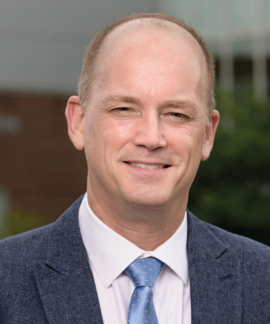
Distinguished Professor
Department of Biological Sciences, College of Science
The Tesmer lab studies the molecular basis of GPCR-mediated signal transduction using techniques such as SAXS, X-ray crystallography, and single particle cryo-EM reconstruction. By determining structures of signaling proteins alone and in complex with their various targets, trainees discover mechanisms underlying signal transduction, explain how diseases result from dysfunctional regulation, and explore how to leverage this information to engender new therapeutic approaches. The lab also investigates the structure and function of LCAT, a key enzyme in reverse cholesterol transport, with the goal of understanding its activation by HDL particles and its design into a therapy for human somatic disease and acute coronary syndrome. Since its inception 1999, the lab has trained 21 Ph.D./M.S. students (including 1 URM).
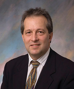
Professor
Department of Chemistry, College of Science
Dr. Thompson studies lipid-based systems to include light and acid-labile liposomes, oriented insertion of membrane proteins in supported lipid membranes, interfacial protein crystallization, affinity capture materials for cryo-EM, and understanding the intracellular mechanism of cargo release from transfection materials. He has published over 140 papers in the areas of bioresponsive material development for small molecule and nucleic acid delivery, surface modification for accelerated protein structure determination, and supported membrane protein sensors for drug discovery.
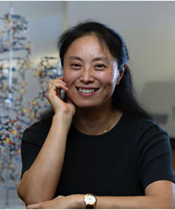
Distinguished Professor
Department of Medicinal Chemistry and Molecular Pharmacology, College of Pharmacy
Dr. Yang's research is focused on structures and functions of cancer-specific DNA molecular targets and structure-based rational design of new anticancer drugs, with the goal of combining the potency of DNA-interactive compounds with the selectivity of molecular-targeted cancer therapeutics. Yang's group works on several DNA molecular targets for cancer therapeutics, including DNA secondary structures such as G-quadruplexes, and elucidates biological functions and molecular interactions using small molecules and proteins. A second program involves DNA bis-intercalating drugs that inhibit DNA binding by specific transcription factors. Trainees use a variety of biophysical and biochemical methods for characterization, in particular high-field NMR spectroscopy.